
Multi-Organ Research Reference
Triple-Organ Protection: Seven Compounds Studied Across Brain, Kidney, and Heart
The brain, kidneys, and heart are the body's most metabolically expensive tissues, and they degrade along the same sequence: perfusion drops, reactive oxygen species outpace endogenous antioxidant capacity, and inflammatory signaling amplifies the original insult. This reference summarizes seven research compounds studied against that shared sequence — organized by mechanism rather than by organ.
ROS
Oxidative load is the shared failure mode across all three organ systems
Cardiolipin
SS-31 binds the inner mitochondrial membrane to preserve cristae architecture
SIRT1/3
NAD+ availability gates sirtuin-mediated cellular stress resistance
Overview
Why the brain, kidney, and heart share a failure mode
The brain, kidneys, and heart consume a disproportionate share of resting energy relative to their mass. All three depend on dense microvascular networks to deliver oxygen at the rate their mitochondria are able to consume it, and none of the three holds a meaningful energy reserve.
That shared dependency produces a shared vulnerability. When perfusion falls or electron transport becomes inefficient, each organ degrades along the same sequence: reactive oxygen species (ROS) accumulate faster than endogenous antioxidant systems neutralize them, endothelial function deteriorates, and inflammatory signaling amplifies the initial insult into permanent tissue loss.
Research into multi-organ protection follows that common sequence rather than treating the three organs as separate problems. The compounds below are studied across neural, renal, and cardiac models precisely because the mechanisms they act on — glutathione status, cardiolipin integrity, NAD+ availability, melanocortin signaling — are not organ-specific.
Mechanism
Three levers the literature returns to
Across the neural, renal, and cardiac literature, protective research converges on three intervention points. Each compound in this reference acts on one or more of them.
- Microvascular and endothelial preservation — maintaining nitric oxide bioavailability and barrier integrity so perfusion survives the insult.
- Mitochondrial bioenergetics — preserving cristae architecture, electron-transport coupling, and ATP output, rather than only scavenging the ROS that inefficiency produces.
- Cellular stress resistance — glutathione status, sirtuin activity, and inflammatory tone, which together determine whether a stressed cell recovers or commits to apoptosis or ferroptosis.
Antioxidant
Glutathione (GSH)
Glutathione is the principal endogenous antioxidant — a tripeptide held at millimolar concentration inside most cells, where it acts as a direct ROS scavenger, a conjugation substrate for xenobiotic clearance, and the cofactor that allows glutathione peroxidase 4 to suppress ferroptosis.
The science: GSH depletion is a common early event across all three organ systems. Where the pool holds, lipid peroxidation stays contained; where it collapses, membrane phospholipids oxidize and cells commit to ferroptotic death.
Brain: endogenous GSH homeostasis has been characterized as preventing excitotoxic cell death and limiting regional oxidative injury in neurodegenerative models (Monks, 1999).
Kidney: the kidney extracts circulating glutathione directly from plasma, and this renal uptake is reported to protect tubular epithelial cells against ischemic and nephrotoxic damage (Lash, 2024).
Heart: enhancing the glutathione system is reported to limit myocardial lipid peroxidation and prevent ferroptotic cell death during ischemic events (Tan et al., 2023).
Precursor
N-Acetylcysteine (NAC)
NAC supplies cysteine, the rate-limiting substrate for glutathione synthesis. Where glutathione is the effector, NAC is the upstream input — which is why the two appear together throughout the protection literature.
The science: beyond replenishing the GSH pool, NAC scavenges hydroxyl radicals directly and is reported to promote eNOS-mediated vasodilation, giving it a vascular mechanism independent of its antioxidant role.
Brain: reported to suppress ischemic brain injury and preserve local perfusion following transient cerebral ischemia (Tenório et al., 2021).
Kidney: downregulates transforming growth factor beta-1 (TGF-β1) and has been studied against renal ischemia–reperfusion injury and contrast-induced nephropathy (Tan et al., 2022; Watanabe et al., 2021).
Heart: reported to reduce oxidative stress-induced cardiac injury and improve functional recovery following cardiac surgery and ischemic insult (Tenório et al., 2021).
Note: NAC is not currently stocked in the Revitalized catalog. It appears in this reference because the glutathione literature is difficult to read without it.
Mitochondrial
SS-31 (Elamipretide)
SS-31 is a mitochondria-targeted tetrapeptide that binds selectively to cardiolipin, a phospholipid found almost exclusively on the inner mitochondrial membrane.
The science: rather than neutralizing ROS after they form, SS-31 is characterized as acting on the source. Stabilizing cardiolipin holds cristae architecture in place, keeps electron transport coupled, and reduces the superoxide leak that uncoupling produces.
Brain: reported to protect against blood–brain barrier disruption and neuronal apoptosis in cerebral ischemia models (Birk et al., 2013).
Kidney: reported to preserve podocyte structure and attenuate glomerulosclerosis and tubular injury in aging and ischemic kidney models (Sweetwyne et al., 2017).
Heart: reported to restore mitochondrial ATP production and limit cardiac remodeling following myocardial ischemia–reperfusion (Birk et al., 2013).
Anti-inflammatory
KPV (α-MSH 11–13)
KPV is the C-terminal tripeptide of α-melanocyte-stimulating hormone — lysine-proline-valine, the smallest fragment reported to retain the parent hormone's anti-inflammatory activity without its pigmentary effects.
The science: KPV is studied as a suppressor of NF-κB activation, reducing downstream pro-inflammatory cytokine production. Because inflammation is the amplifier that converts a survivable ischemic insult into permanent tissue loss, KPV sits at a different point in the sequence than the antioxidants above.
Brain: reported to mitigate neuroinflammation and microglial activation in central nervous system injury models (Hao et al., 2024).
Kidney: reported to reduce inflammatory infiltration and tissue damage during acute ischemic renal insults (Hao et al., 2024).
Heart: reported to promote tissue repair and suppress adverse inflammatory remodeling following myocardial infarction (Hao et al., 2024).
Cofactor
Nicotinamide Adenine Dinucleotide (NAD+)
NAD+ is the cofactor gating two families of stress-response enzymes: the sirtuins — SIRT1 and SIRT3 in particular — and the PARP DNA-repair enzymes.
The science: the NAD+ pool declines with age and falls sharply under ischemic stress, and PARP activation during DNA damage consumes it further. The literature treats restoring that pool as a way of keeping sirtuin-mediated survival signaling available at the moment the cell most needs it.
Brain: reported to restore cellular ATP levels and protect neurons against ischemic stroke and age-related neurodegeneration (Conlon, 2021).
Kidney: reported to prevent acute kidney injury by restoring renal tubular bioenergetics and sirtuin pathway activity (Hershberger et al., 2017).
Heart: reported to mitigate ischemic heart injury and blunt pathological hypertrophy through sirtuin-mediated cardioprotective signaling (Hershberger et al., 2017).
Substrate transport
L-Carnitine
L-Carnitine shuttles long-chain fatty acids across the inner mitochondrial membrane for β-oxidation. Without adequate carnitine the substrate never reaches the enzymes that oxidize it.
The science: the heart draws the majority of its ATP from fatty-acid oxidation, and the literature describes carnitine depletion as producing metabolic inflexibility — a state in which a stressed cell cannot switch fuel sources, and energy output falls independently of oxygen supply.
Brain: reported to maintain mitochondrial membrane potential and protect cortical neurons against amyloid toxicity and metabolic failure (Virmani & Cirulli, 2022).
Kidney: reported to reduce tubular cell damage in models of contrast-induced nephropathy and diabetic renal injury (Panova & Tatikolov, 2023).
Heart: reported to enhance myocardial energy production, reduce ischemic cardiac damage, and support post-infarction recovery (Virmani & Cirulli, 2022).
Ion channel
PE-22-28
PE-22-28 is a shortened spadin-derived peptide and a potent antagonist of the TREK-1 two-pore-domain potassium channel, reported with an IC₅₀ near 0.12 nM.
The science: TREK-1 blockade sits outside the antioxidant and bioenergetic mechanisms above — it is a signaling intervention. The channel regulates neuronal excitability and contributes to neurovascular coupling and vascular smooth-muscle tone.
Brain: reported to stimulate hippocampal neurogenesis, exert neuroprotective effects, and aid neuronal recovery following stroke (Djillani et al., 2017).
Kidney and heart: direct renal and cardiac endpoints are not established for this compound. Active investigation centers on TREK-1 regulation within neurovascular coupling and systemic vascular smooth-muscle tone, with broader microvascular implications still at the preclinical stage. It is included here for mechanistic completeness, not for parity of evidence with the six compounds above.
Where they sit in our catalog
Six of the seven are stocked
Glutathione, SS-31, KPV, NAD+, L-Carnitine, and PE-22-28 are available as research compounds. NAC is referenced for mechanistic context only and is not part of the catalog.
References
Selected literature
Birk, A. V., et al. (2013). The mitochondrial-targeted compound SS-31 re-energizes ischemic mitochondria by interacting with cardiolipin. Journal of the American Society of Nephrology, 24(8), 1250–1261.
Conlon, N. J. (2021). The role of NAD+ in regenerative medicine. Plastic and Reconstructive Surgery, 150(4S), 41S–48S.
Djillani, A., et al. (2017). Shortened spadin analogs display better TREK-1 inhibition, in vivo stability and antidepressant activity. Frontiers in Pharmacology, 8, 643.
Hao, Z.-W., et al. (2024). Bioactive peptides and proteins for tissue repair: microenvironment modulation, rational delivery, and clinical potential. Military Medical Research, 11(1).
Hershberger, K. A., et al. (2017). Role of NAD+ and mitochondrial sirtuins in cardiac and renal diseases. Nature Reviews Nephrology, 13(4), 213–225.
Lash, L. H. (2024). Renal glutathione: Dual roles as antioxidant protector and bioactivation promoter. Biochemical Pharmacology, 228, 116181.
Monks, T. (1999). Symposium overview: the role of glutathione in neuroprotection and neurotoxicity. Toxicological Sciences, 51(2), 161–177.
Panova, I. G., & Tatikolov, A. S. (2023). Endogenous and exogenous antioxidants as agents preventing the negative effects of contrast media. Pharmaceuticals, 16(8), 1077.
Sweetwyne, M. T., et al. (2017). The mitochondrial-targeted peptide, SS-31, improves glomerular architecture in mice of advanced age. Kidney International, 91(5), 1126–1145.
Tan, M., et al. (2023). Glutathione system enhancement for cardiac protection: pharmacological options against oxidative stress and ferroptosis. Cell Death & Disease, 14(2).
Tan, Y. K., et al. (2022). N-acetylcysteine's renoprotective effect in cardiac surgery: A systematic review and meta-analysis. Annals of Thoracic and Cardiovascular Surgery, 28(2), 138–145.
Tenório, M. C. D. S., et al. (2021). N-acetylcysteine (NAC): Impacts on human health. Antioxidants, 10(6), 967.
Virmani, M. A., & Cirulli, M. (2022). The role of L-carnitine in mitochondria, prevention of metabolic inflexibility and disease initiation. International Journal of Molecular Sciences, 23(5), 2717.
Watanabe, M., et al. (2021). Renoprotective effect of N-acetylcysteine depends upon the severity of the ischemia reperfusion injury. Brazilian Journal of Medical and Biological Research, 54(9).
Safety & disclaimers
Research use only — not medical advice
This article is for educational purposes only. It summarizes published research and does not constitute medical advice, diagnosis, or treatment. The findings described are observations from the cited literature, not guidance for personal use.
All Revitalized research compounds are sold for research use only. Not for human consumption, diagnostic, or therapeutic use. Standard institutional biosafety guidelines apply.
Related Compounds
Continue Researching This Category
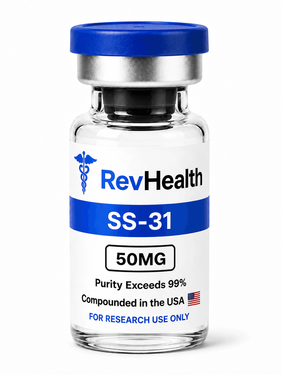
SS-31 50MG
Mitochondrial-targeted antioxidant peptide
from $75
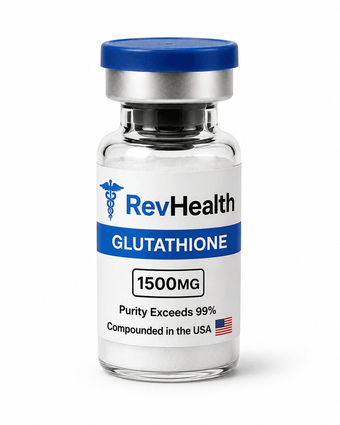
GLUTATHIONE 1500MG
Master antioxidant for detox and skin research
from $75
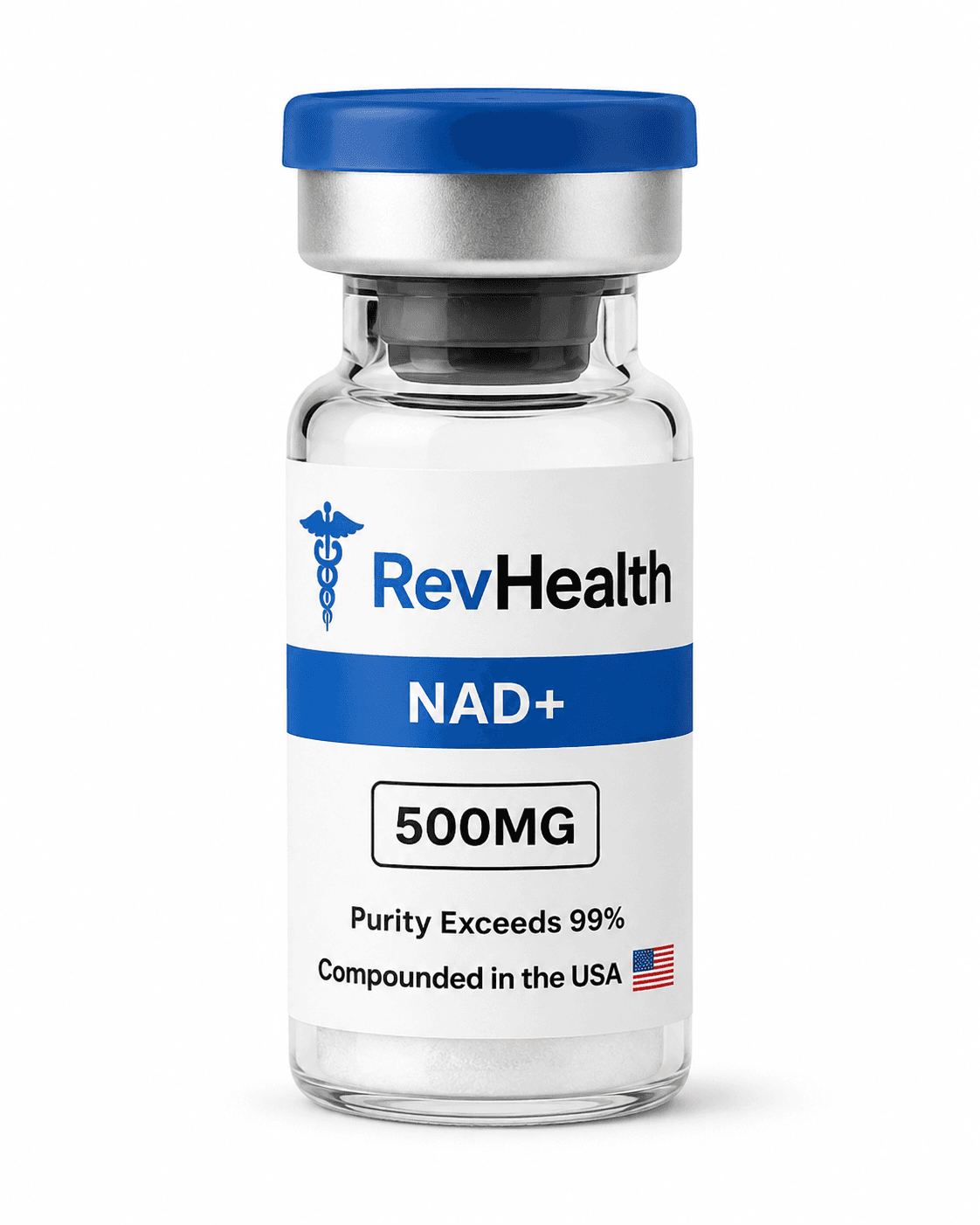
NAD+ 500MG
Cellular energy and longevity research compound
from $70
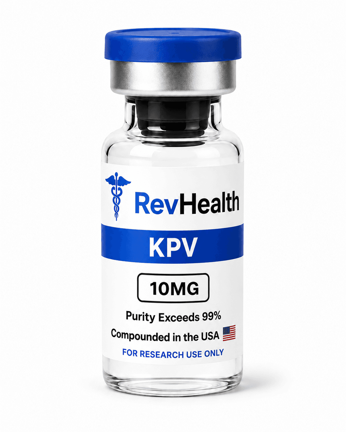
KPV 10MG
Anti-inflammatory tripeptide for gut health research
from $55
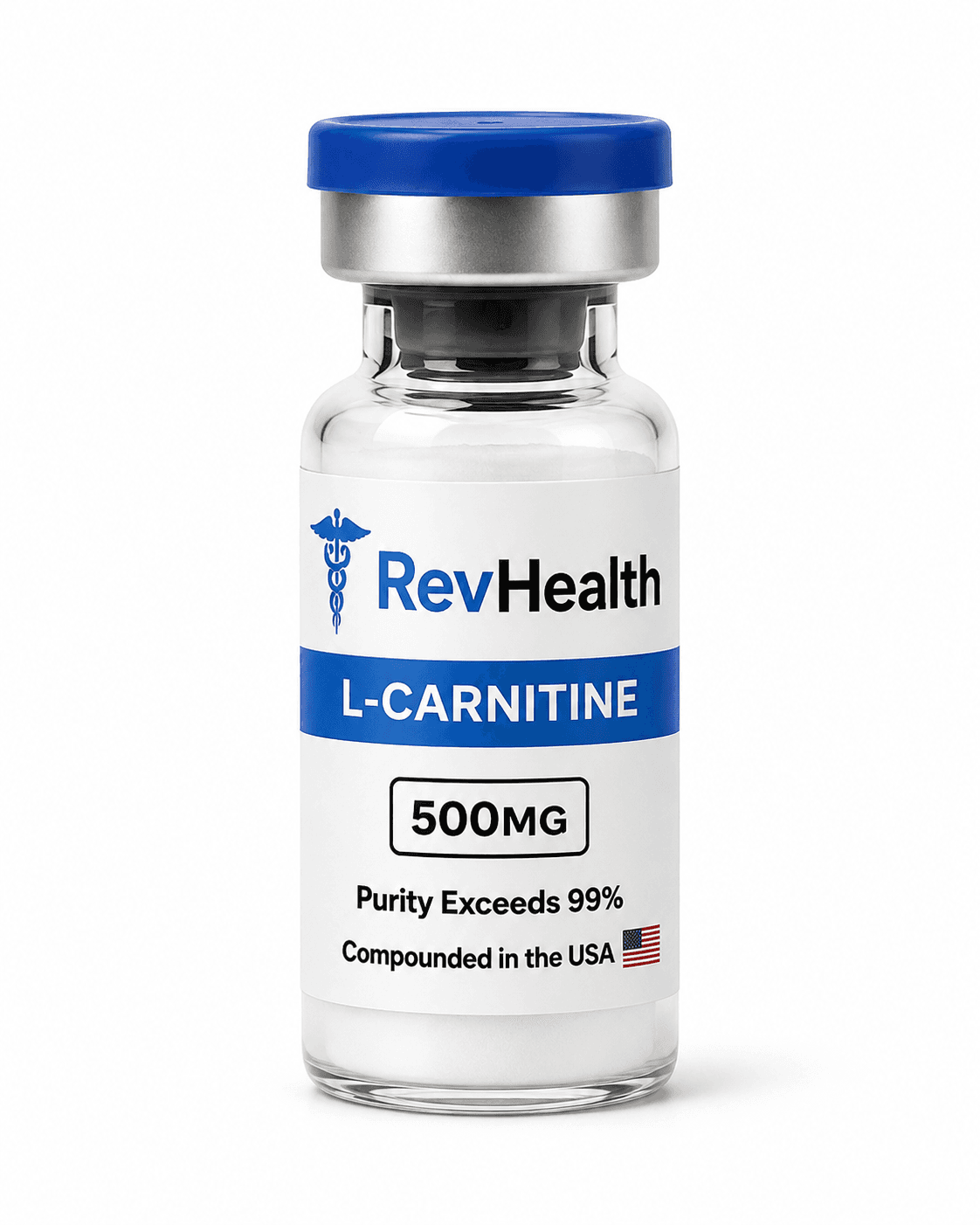
L-CARNITINE
Fat metabolism and energy production compound
from $70
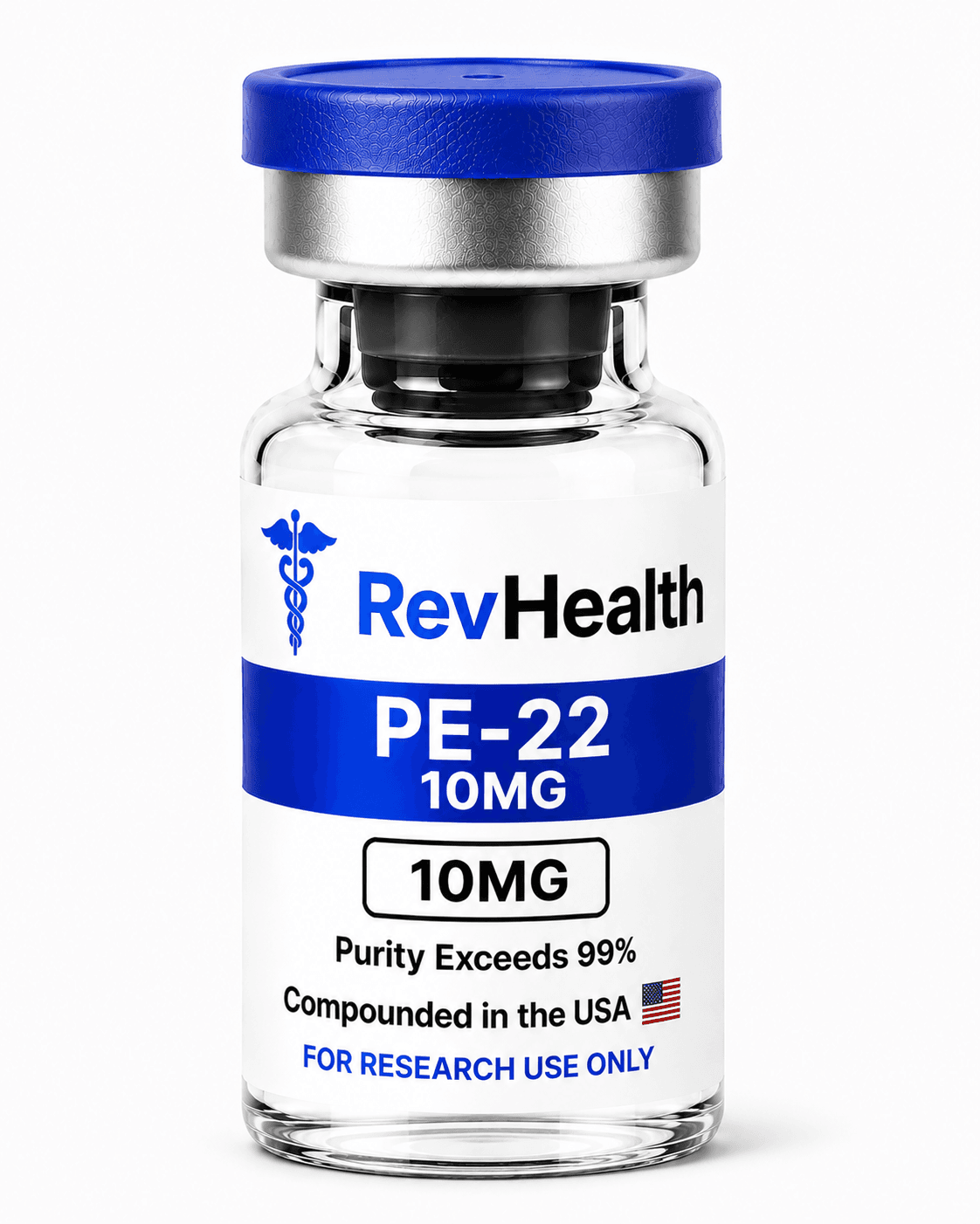
PE-228
Sortilin-derived neuropeptide for TREK-1 channel research
from $75
Continue From This Update
Explore the research catalog behind these topics.
Browse verified research compounds with third-party analytical documentation and category-specific product pages.
Shop Research PeptidesResearch use only. This article is educational and does not constitute medical advice. Products referenced on Revitalized Health are not intended for human or veterinary use, diagnostic use, therapeutic use, or consumption.